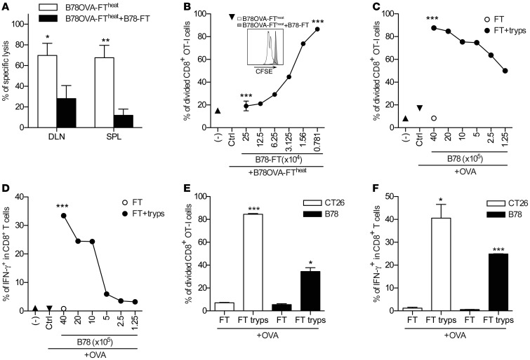
Figure 2. Primary FT necrotic cells abort cross-priming of CD8+ T cells.
(A) B78OVA-FTheat cells were mixed with B78-FT cells at a 1:8 ratio and injected into mice on days 0 and 2, so that each mouse was vaccinated with a total of 2 × 10⁶ B78OVA cells. Ag-specific immune responses were analyzed on day 7. Data are mean ± SEM of 2 pooled experiments (n = 3 per group). (B) SF of 2.5 × 10⁵ B78OVA-FTheat cells was cultured in vitro with CFSE-labeled OT-I splenocytes, and titrated amount of SF from B78-FT was added. Proliferation of CD8+ T cells was analyzed after 48 hours. Data are representative of at least 3 independent experiments. Inset shows proliferation of OVA-specific CFSE-labeled CD8+ T cells cultured with B78OVA-FTheat or B78OVA-FTheat +B78-FT cells. (C and D) SF from B78-FT cells was trypsin digested and added at different dilutions to CFSE-labeled OT-I cells in the presence of 20 μg/ml OVA. Proliferation (C) and IFN-γ expression (D) of CD8+ T cells were analyzed 48 hours later. Data are representative of at least 3 experiments. (E and F) Trypsin-digested SF from CT26-FT or B78-FT cells were added to CFSE-labeled OT-I splenocytes in the presence of 20 μg/ml OVA, and Ag-specific proliferation (E) and IFN-γ expression (F) was tested. Data are representative of at least 3 experiments. *P < 0.05, **P < 0.01, ***P < 0.001, Student’s t test (A and C–F) or 1-way ANOVA with Dunnett’s multiple-comparison test (B).

