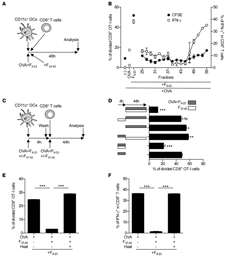
Figure 4. F37–45 characterization.
(A and B) Chromatographic fractions (see Figure 2E) were added to CFSE-labeled OT-I splenocytes in the presence of 20 μg/ml OVA and F9–23. Proliferation and IFN-γ expression of CD8+ T cells was analyzed after 48 hours. Data show 1 representative of 2 independent experiments. (C and D) Splenic CD11c+ DCs (5 × 10⁴) were preincubated with F9–23 and 20 μg/ml OVA with or without F37–45 for 4 hours. DCs were then washed, and purified CFSE-labeled OT-I CD8+ T cells (1 × 10⁵) were added and incubated for 44 hours in the presence of F37–45 and/or F9–23 plus OVA, as indicated. Proliferation of Ag-specific CD8+ T cells was determined. (E and F) 20 μg/ml OVA protein was incubated with F37–45 overnight at 37°C and subsequently heated at 70°C for 1 hour as indicated. Samples were added to the CFSE-labeled OT-I cells in the presence of F9–23, and CD8+ T cell activation was determined after 48 hours. Results are representative of 3 independent experiments. *P < 0.05, **P < 0.01, ***P < 0.001, 1-way ANOVA with Dunnett’s multiple-comparison test (D) or Student’s t test (E and F).

