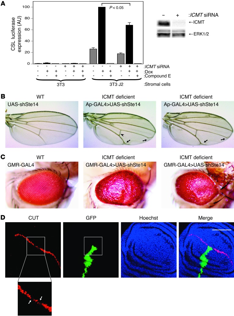
Figure 8. ICMT deficiency inhibits Notch1 signaling.
(A) Notch signaling in mammalian cells quantified with a CSL firefly luciferase reporter. Shown are the ratios of CSL firefly/CMV Renilla in U2OS cells containing doxycycline-inducible FLAG-Notch1-GFP. Cells were cocultured with the indicated “stromal cells” in the presence or absence of doxycycline (Dox) and siRNA targeting ICMT or a nontargeting control. The stromal cells were NIH3T3 (3T3) with or without expression of the Notch1 ligand Jagged-2 (J2). The γ-secretase inhibitor compound E was included as a control and completely blocked Notch signaling. All ratios were normalized to the maximum in each experiment. Data shown are mean ± SEM, n = 3. Panel on right shows a representative knockdown by immunoblotting for ICMT and a control (ERK1/2). (B) Wings of D. melanogaster transgenic for UAS-shSte14, a GAL4-responsive hairpin that silences Icmt. GAL4 expression was driven by a wing-specific (Ap-GAL4) promoter. ICMT deficiency in the developing wing phenocopies the terminal vein bifurcation (arrows) and thickened cross-vein (arrowhead) observed in the wings of Delta (Dl) flies deficient for the Notch ligand. (C) GAL4 expression driven with an eye-specific (GMR-GAL4) promoter. The rough eye phenotype is also consistent with that seen in Notch loss-of-function alleles, including Dl. (D) Wing imaginal discs from third instar larvae expressing shSte14 in heat shock–induced clones marked with GFP and stained for CUT, a Notch-dependent gene product. Where the GFP-positive clone intersects the line of CUT staining there is a decrease in the number of CUT-positive cells (enlargement). Scale bar: 50 εm.

