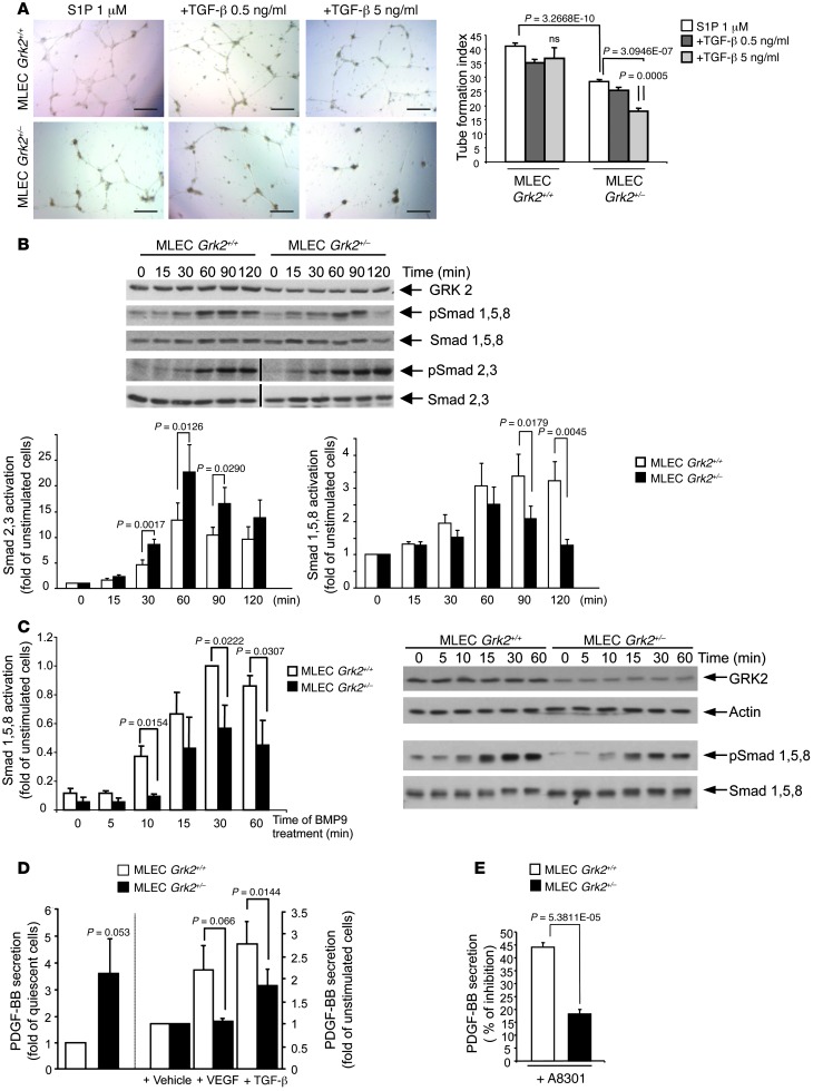
Figure 3. Effect of GRK2 downregulation on TGF-β1 endothelial signaling.
(A) ECs with reduced expression of GRK2 are more sensitive to dose-dependent inhibitory effects of TGF-β1 on tube network formation. In vitro tube formation was monitored in the presence of 1 μM S1P supplemented with or without 0.5 or 5 ng/ml TGF-β1 and quantified as in Figure 2, D and E. Data from 3 independent assays performed in duplicate are shown. Scale bar: 500 μm. (B and C) Proper signaling of Smads downstream of ALK1 and ALK5 receptor activation depends on GRK2 expression. (B) Grk2+/– MLECs display enhanced TGF-β1–triggered activation of Smad2/3 paralleled by a decrease in Smad1/5/8 stimulation. The lanes were run on the same gel but, where indicated by the black vertical line, were noncontiguous. (C) BMP9-induced activation of Smad1/5/8 via ALK1 is reduced in Grk2+/– MLECs. (D) Both basal and angiogenic factor–induced PDGF-BB secretion are altered in Grk2+/– MLECs. Serum-starved cells were incubated for 48 hours in 1% FCS supplemented with or without 50 ng/ml VEGF or 5 ng/ml TGF-β1, and PDGF-BB was determined in the cell-conditioned media as described in Methods. (E) The production of PDGF-BB in Grk2+/– MLECs is less sensitive to ALK5 signaling inhibitors. Media of cells treated with 5 ng/ml TGF-β1 for 48 hours in the presence or absence of the ALK5 inhibitor A8301 (5 μM) was processed for PDGF-BB quantification. In B–E data from 3–4 independent experiments are shown.

