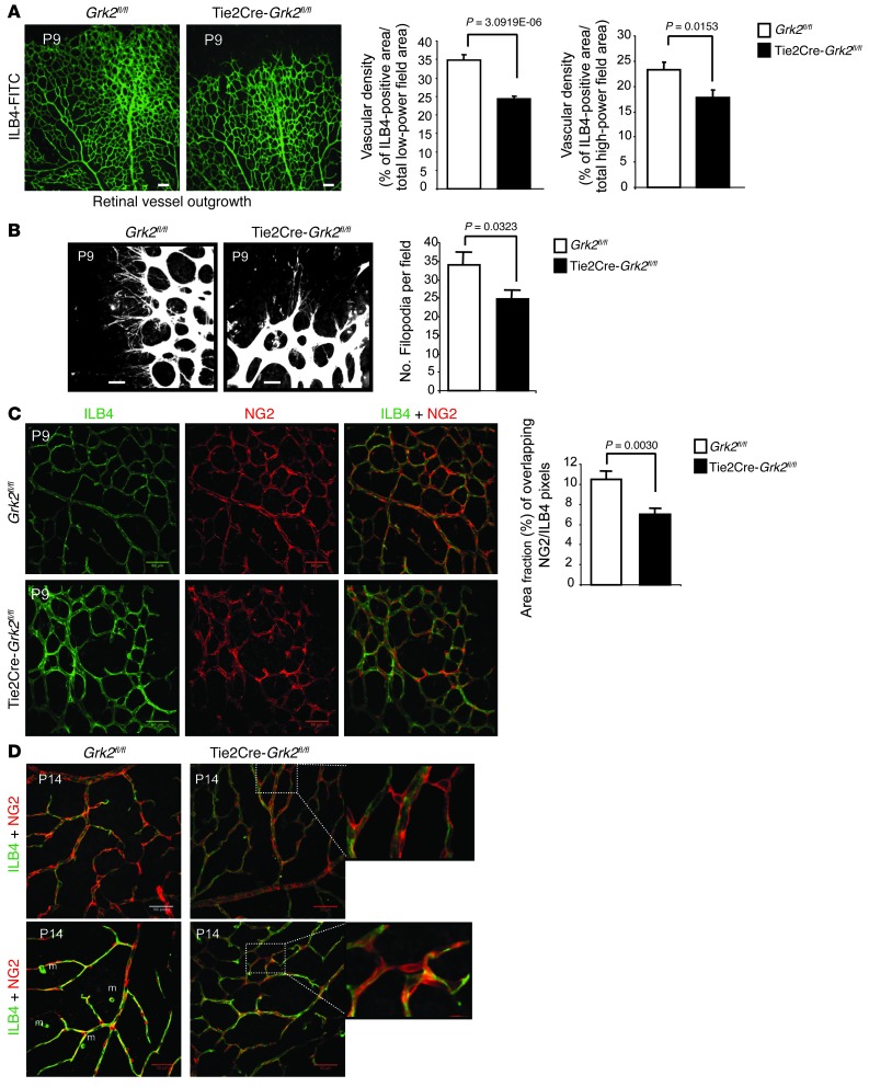
Figure 4. Loss of endothelial GRK2 impairs developmental outgrowth and maturation of the retinal vasculature.
(A and B) Expansion of the primary vascular plexus and endothelial filopodia was reduced in the absence of GRK2. (A) Whole-mount P9 retinas from WT (Grk2fl/fl; n = 5) or Tie2Cre-Grk2fl/fl (n = 5) pups were stained with endothelial FITC-ILB4 marker. The ILB4-positive area was quantified in the entire retinal surface (low magnification) or in proximal regions of the vascular plexus (high magnification) (WT, n = 15; mutant, n = 12) as detailed in Methods. (B) Filopodia of tip cells were counted in high-power fields of 5 WT retinas (n = 17) and 6 Tie2Cre-Grk2fl/fl retinas (n = 18). (C and D) Delayed recruitment of pericytes to retinal ECs and perturbed cell-cell interactions in mutant mice. Whole-mount (C) P9 and (D) P14 retinas of WT (n = 5) and Tie2Cre-Grk2fl/fl (n = 5) mice were double stained with ILB4-FITC and anti-NG2 antibodies, and the corresponding positive areas were quantified (n = 14 and n = 11 images, respectively) as detailed in Methods. (C) Pericyte coverage was expressed as the percentage fraction of colocalizing pericyte- and endothelial-positive areas. (D) Abnormal association of pericytes with endothelial capillaries in the primary vascular plexus of P14 Tie2Cre-Grk2fl/fl retinas. Some pericytes remain dissociated from ECs and make irregular connections between endothelial capillaries (zoomed images). High-magnification fields (n = 12 from retinas of 5 Grk2fl/fl; n = 20 from retinas of 5 Tie2Cre-Grk2fl/fl) were inspected. m, macrophages. Scale bar: 100 μm (A, C, and D); 25 μm (B).

