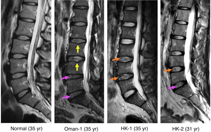
Figure 3. MRI of heterozygous individuals with known mutations in CHST3.
T2 MRI of a normal individual and parents of patients with recessive spondyloepiphyseal dysplasia (OMIM 603799) from Oman (Oman-1) and Hong Kong (HK-1 and HK-2). Disc degeneration with reduced NP intensity (pink arrows), abnormal end-plate (yellow arrows) consistent with Schmorl’s nodes, and irregular darkened NP signals (orange arrows) were observed.

