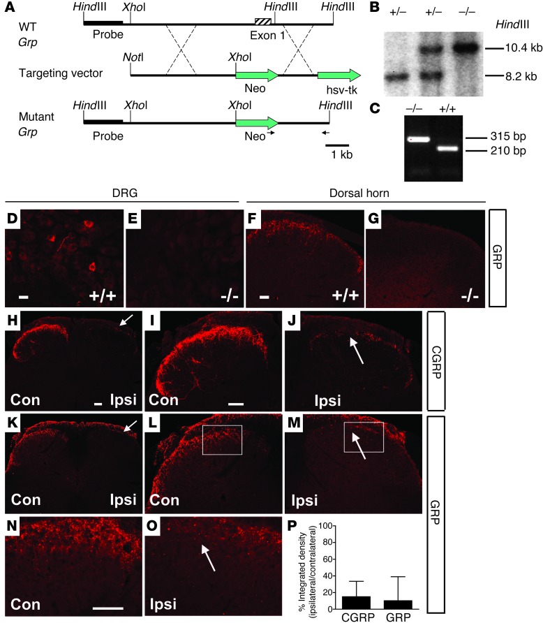
Figure 6. Generation of Grp–/– mice and confirmation of GRP expression in primary sensory neurons.
(A) Targeting strategy for generation of Grp–/– mice. (B and C) Germ line transmission was confirmed by (B) Southern blot and (C) PCR analysis. (D–G) GRP expression in the (D and E) DRGs and (F and G) spinal cords of (D and F) wild-type mice and (E and G) Grp–/– mice. (H–O) Expression of (H–J) CGRP and (K–O) GRP in the lumbar spinal cords of C57BL/6J mice 14 days after unilateral dorsal rhizotomy (L5). On contralateral sides, both (H and I) CGRP+ and (K, L, and N) GRP+ fibers were mainly in the superficial dorsal horn (lamina I, IIo); but on the ipsilateral sides, both (H and J) CGRP and (K, M, and O) GRP staining was lost after unilateral L5 dorsal rhizotomy. Arrows indicate the elimination of staining. n = 3 per group. (P) Quantitation of remaining CGRP+ (15.1%) and GRP+ (10.2%) staining in the L5 superficial dorsal horn after rhizotomy. Boxed areas in L and M are shown at higher magnification in N and O. Scale bar: 10 μm (D and E); 20 μm (F and G); 40 μm (H–O).

