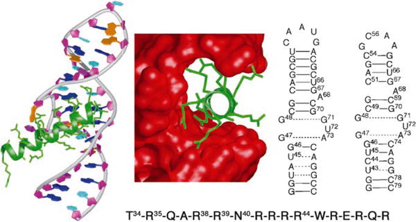
Figure 1.

Left: X-ray structure of Rev (green) bound to HIV-1 RRE IIb (prepared from protein data base (PDB) number: 1ETF); middle: key residues in Rev (green) bound in the major groove of the RRE (red surface); right: secondary structure of RRE IIb Rev binding site, used in EMSA studies and the secondary structure of a modified RRE IIb used for NMR spectroscopic studies; bottom: sequence of the arginine-rich RNA binding domain (Rev34–50) of HIV-1 Rev protein.
