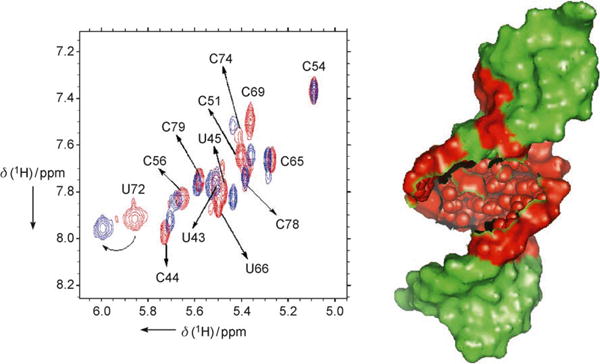
Figure 4.

Left: superimposition of TOCSY spectra of free RRE-RNA in red (sequence shown in Figure 1 top, right) and 1:1 BIV-5:RRE complex in blue. Resonances at or near the purine-rich internal loop are affected by peptide binding, while the rest of the RNA is not. Homonuclear 1H TOCSY and NOESY experiments of the free RNA and BIV-5-RRE complex were recorded at 750 MHz (Bruker DMX-750) at RNA concentrations of 0.5–1.5 mM in D2O at pH 6.6 in phosphate buffer; right: schematic representation of the region of RRE affected by peptide binding (red) and the region that remained unaffected (green).
