
Figure 1.
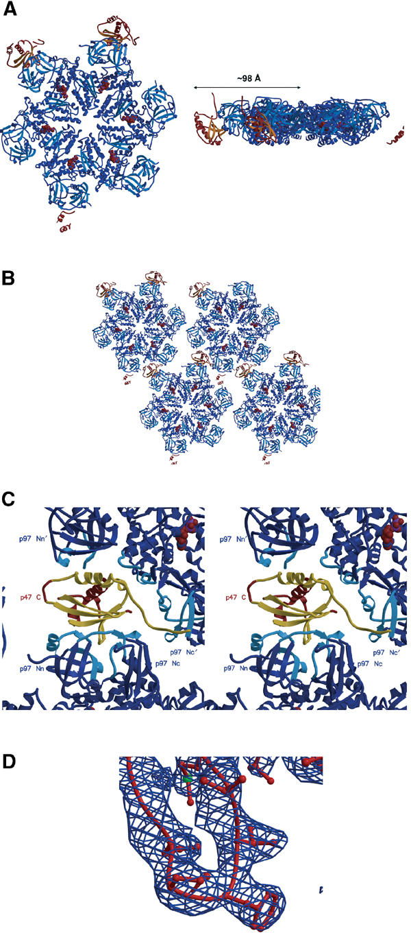
Overview of the p97 ND1–p47 C structure. (A) Ribbon representation of the p97 ND1 (blue)–p47 C (red/orange) crystal structure (top and side views). The radius of the complex structure is indicated. The disconnected secondary structure at the bottom (and right) of the p97 hexamer corresponds to parts of a third p47 C molecule we observe in the density maps. (B) Crystal packing arrangement of the p97 ND1–p47 C complex in one plane as viewed down the crystallographic 65 screw axis. (C) Stereo close-up view of one p47 C molecule (in red/gold) in the crystal lattice. The N domains from two ND1 hexamers (blue) are labelled. The ND1 secondary structure elements from the two hexamers that form the p47 C interface are depicted in light blue (‘binary' complex of p47 UBX with p97 Nn and p97 Nc; additional lattice contact between the N-terminal extended chain of p47 C with the p97' hexamer). p47 C secondary structure elements that interact with ND1 are depicted in gold. (D) Electron density (2Fo-Fc map contoured at 1.2σ) of the p47 C S3/S4 loop region.