
Figure 3.
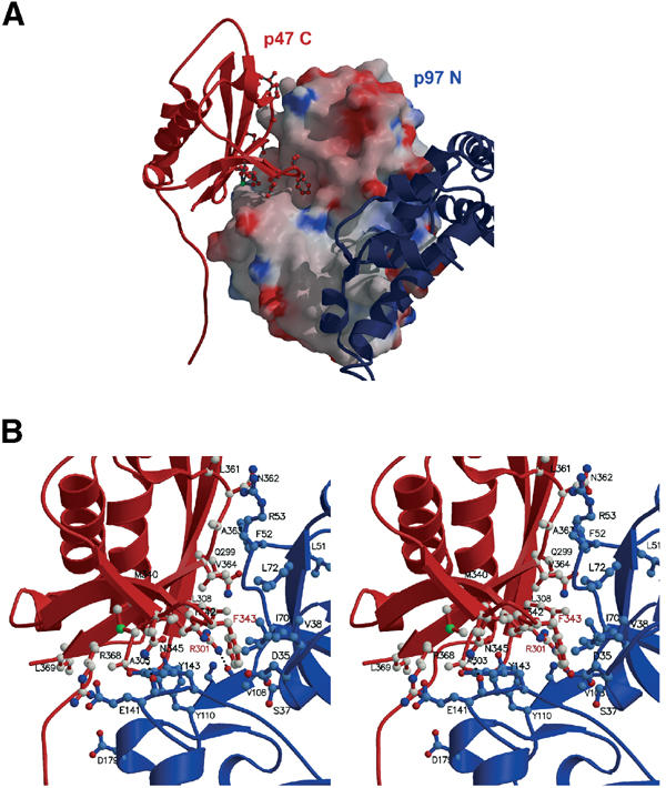
Binding of p47 C to p97 N domain. (A) Electrostatic surface representation of p97 N domain interacting with p47 C (ribbon coloured red). Key residues of p47 C at the N-domain interface are shown in ball-and-stick representation. D1 is depicted in blue. (B) Detailed view of specific interactions at the p47 C–p97 N interface. p47 C is shown as a ribbon representation (red) with key residues in ball-and-stick, p97 N is depicted in blue. Hydrogen-bonding interactions (p47 C Arg301NH2 and p97 N Val108O as well as p47 C Asn345ND2 and p97 N Glu141O) are indicated by a dotted line. Key residues conserved within UBX domains are labelled in red.