
Figure 4.
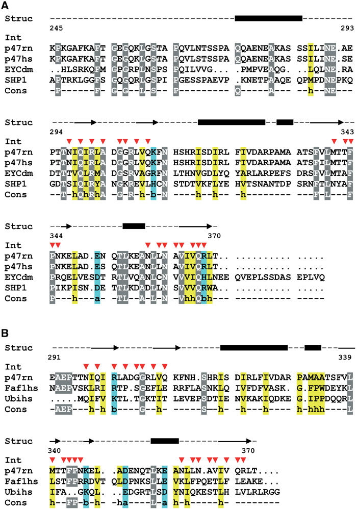
Sequence alignments showing p97–p47 interacting residues. (A) Sequence alignment of p47 homologues (rat p47: accession code tr∣O35987∣, human p47: accession code tr∣Q9UNZ2∣, Drosophila EYC: accession code tr∣Q9U9C9∣ and yeast SHP1: accession code sp∣P34223∣) starting at residue 245 of rat p47. (B) Structure-based sequence alignment of p47 and FAF1 UBX domains and ubiquitin. Residues located at the interface with p97 N in the ‘binary' complex are marked with a red arrow. Secondary structure elements of p47 C are shown above the alignment. Conserved residues are highlighted in grey, ‘h' stands for hydrophobic (yellow), ‘b' for basic and ‘a' for acidic residues (blue).