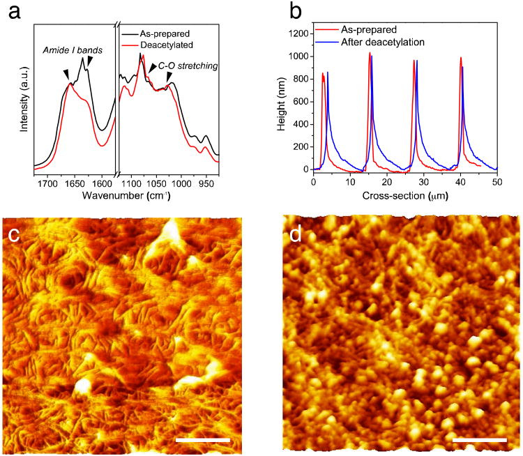
Fig. 3.
Effect of post-treatment of chitin substrates for biological testing (deacetylation in NaOH, fibronectin treatment). (a) FT-IR spectra, (b) Cross-sectional height profiles of the chitin substrates obtained from the G2 grating mold, (c, d) Morphology of chitin nanofiber films after deacetylation in NaOH and fibronectin treatment, respectively (All scale bars = 200 nm).

