Fig. 5.
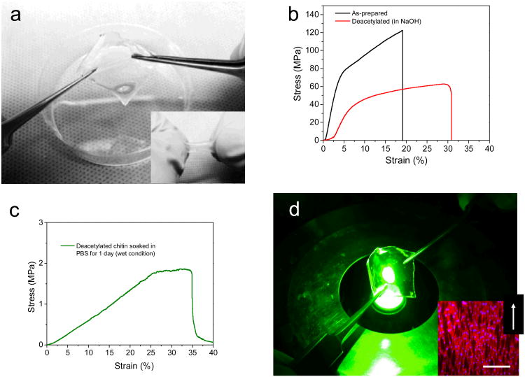
Cell seeded free-standing flexible chitin substrates, (a) Stretching and Rolling (insert) flexibility the free-standing micropatterned chitin substrates (G2) seeded with 3T3 fibroblast (b) Mechanical properties of micropatterned free standing chitin substrates before and after deacetylation to 30%, (c) mechanical properties of the 30% deacetylated chitin nanofiber substrates after immersion in PBS for 1 day (measured wet) (d) Chitin nanofiber substrates are transparent and afford optical inspection, insert Fluorescence images of actin cytoskeleton of the cells on G2 showing the entire covering and alignment of cells within the direction of the patterned features. White arrow on the right corner of the images indicates the direction of the patterns. Scale bar 100 μm.

