
Figure 4.
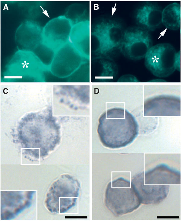
Cholesterol extraction disrupts compartmentalization of NADPH oxidase activity on the plasma membrane. Control (A, C) or cholesterol-extracted (B, D) Ra2 microglia cells were stained with filipin (A, B) to illustrate the efficiency of cholesterol removal. Asterisks denote intracellular stores of cholesterol (which are partially obscured in (A) due to the intense plasma membrane staining of control cells). Arrows point to plasma membrane staining, which is hardly visible in (B). (C, D) Cells were stimulated with PMA in the presence of NBT. Note the pronounced patchy distribution of the precipitation product in the control cells (C), and that cholesterol extraction causes the deposit to become diffusely distributed on the plasma membrane (D). Framed boxes are shown at 2 × magnification. Bars (A–D), 10 μm.