
Figure 7.
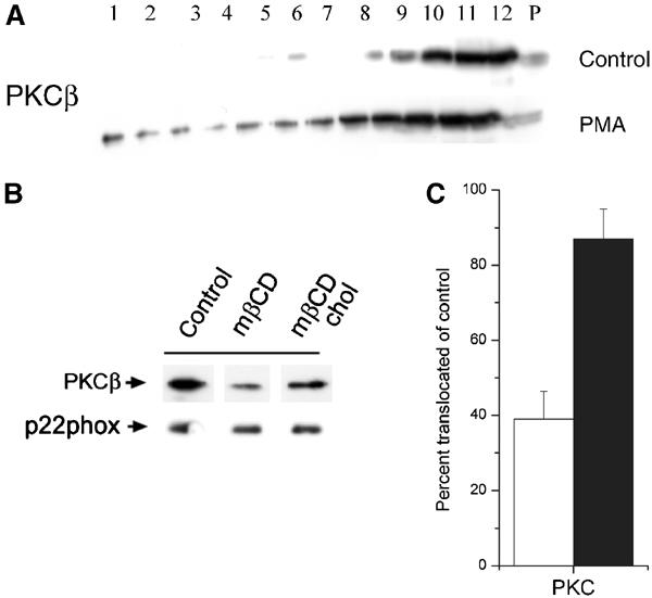
PKC membrane translocation is inhibited after mβCD-mediated cholesterol extraction. (A) Brij-58 lysates of control or PMA-stimulated HL60 cells were centrifuged to equilibrium in a sucrose gradient, and then analyzed by Western blotting using antibodies to PKCβ. (B) HL60 cells were subjected to cholesterol extraction (mβCD), in some cases followed by cholesterol replenishment (mβCD chol), before stimulation with PMA, and translocation of PKCβ to the particulate membrane fraction analyzed by Western blotting. Equal aliquots were also analyzed for p22phox as loading control. (C) The graph represents mean and s.e. of three independent experiments performed as in (B). The results are expressed as percent translocation in cholesterol-extracted (empty bars) or cholesterol-replenished (filled bars) cells relative to control cells.