
Figure 1.
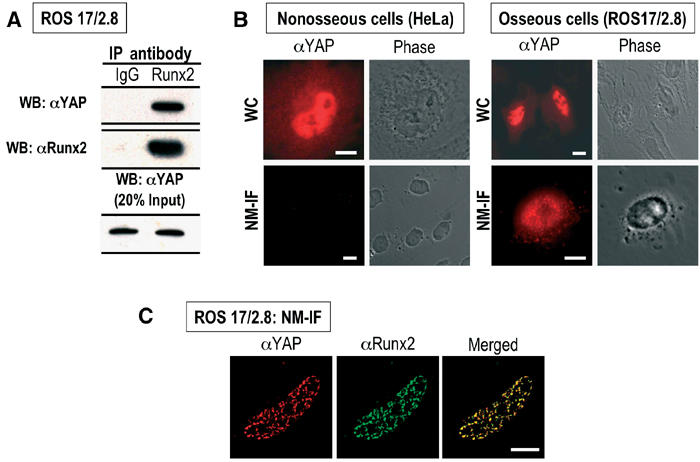
Endogenous YAP and Runx2 proteins interact in vivo and co-localize in situ in osseous cells. (A) Endogenous Runx2 was immunoprecipitated from ROS 17/2.8 cells with a rabbit polyclonal antibody (1:2000) raised against the Runx2 C-terminus (Zhang et al, 2000). A rabbit polyclonal antibody was used to detect endogenous YAP (top panel). Normal goat IgG was used as a control. The middle panel shows efficient immunoprecipitation of endogenous Runx2. The bottom panel shows the expression of endogenous YAP in ROS 17/2.8 cells (20% of total input). (B) In situ immunofluorescence of whole cell (WC) and nuclear matrix-intermediate filament (NM-IF) preparations was performed to assess the nucleo-cytoplasmic distribution and subnuclear localization of endogenous YAP in nonosseous (HeLa) and osseous (ROS 17/2.8) cells. YAP is predominantly nuclear in both HeLa and ROS 17/2.8 cells (top panels), but is only associated with the nuclear matrix ROS 17/2.8 cells (bottom panels). (C) Same as (B), using deconvoluted images. The merged image reveals that endogenous YAP resides in Runx2 containing subnuclear foci in ROS 17/2.8 cells (bar=10 μm).