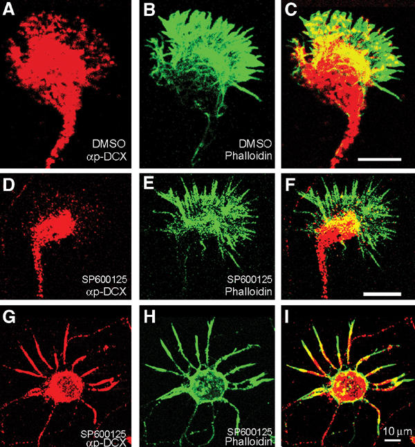
Figure 7.

p-DCX intracellular localization. (A) Rat primary hippocampal growth cone stained with anti-p-DCX, T331, S334 (red) and (B) phalloidin-FITC (green); note the high degree of overlap (C) (yellow). (D–I) Growth cones treated with a specific JNK inhibitor and then stained with anti-p-DCX, T331, S334 (D), or T321 (G) (red) and phalloidin-FITC (green, E, H); note the reduced overlap (F, I, in comparison to C).
