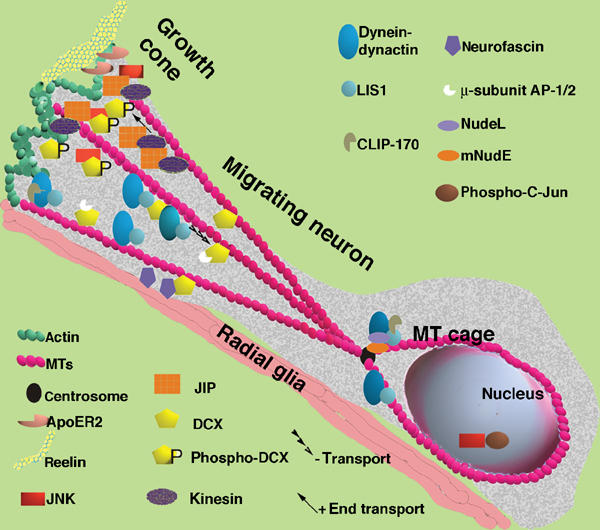
Figure 9.

Model of a neuron migrating along radial glia. This model is based on earlier models (Morris et al, 1998; Feng and Walsh, 2001; Gupta et al, 2002; Hatten, 2002) and incorporates the finding and hypotheses derived from this paper. The migrating neuron has an elongated structure with a growth cone. There is more p-DCX located in the growth cone where it interacts with JNK and JIP. JIP is mobilized there by kinesin. JIP also interacts with ApoER2 that binds to the extracellular matrix protein reelin, and to kinesin that is a plus-end directed motor. DCX also interacts with the membranal protein neurofascin, and with the μ-subunits of the AP-1/2 complexes. At the MT plus-end tips we can also find CLIP-170, which recruits LIS1, and the dynein–dynactin complex. The dynein–dynactin retrograde motor is recruited to MTs with LIS1 and DCX followed by enhanced activity of this motor. Nucleokinesis is assisted by the activity of the dynein motor that is associated with the MT cage and the centrosome (there also mNudE and NudeL can be found). Within the nucleus, the transcription factor c-Jun is phosphorylated by JNK. The activity of JNK may thereby indirectly regulate the differential activities of kinesin and dynein.
