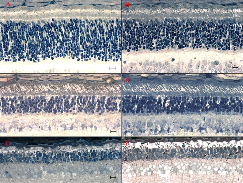
Figure 3.
Light microscopic retinal morphology in crd2-affected dogs exhibiting relative preservation or enhanced development at different ages and between different retinal quadrants. (A, C, E) Superior quadrant (central region, ∼midway from optic nervehead to ora serrata). (B, D, F) Temporal quadrant (central region, ∼midway from optic nervehead to ora serrata). (A, B) Retinas from 3-week-old crd2-affected dogs. Compare also with Figure 2H. Although the ONL is significantly thicker in the superior quadrant (A), IS and OS development is distinctly better preserved in the temporal quadrant (B). (C, D) Retinas from 12-week-old crd2-affected dogs. Compare also to Figure 2I. Preservation of the OS is noticeably better in the temporal quadrant (D), where the IS are more tightly organized and more cones are present, than in the central region (C) with its relatively sparse OSL and disorganized ISL. (E, F) Retinas from 34-week-old crd2-affected dogs. At this age the differences between the central (E) and temporal (F) quadrants are less noticeable, with more severe degenerative changes across the retina. Scale bars: 10 μM.

