
Figure 3. Comparison of 1399R and 1399H rVWF collagen binding.
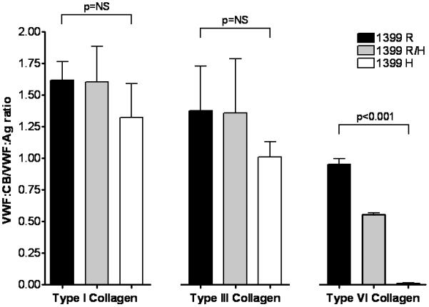
Recombinant full-length VWF was expressed in HEK293T cells using 100% wild-type DNA (1399R, shown in black), 100% variant DNA (1399H, shown in white), or a mixture of 50% of each DNA (1399R/H, shown in gray). Supernatants were analyzed for VWF:Ag and VWF:CB with types I, III, and VI collagen. Graphed here is the ratio of VWF:CB/VWF:Ag for each construct with each type of collagen. Data represent a minimum of 3 separate transfections, and are given as the mean + 1 SD.