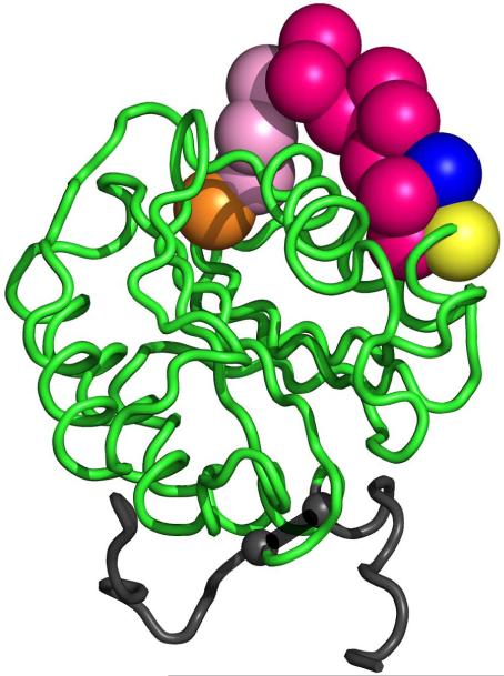
Figure 4. Location of type VI collagen binding mutations in the crystal structure of the A1 domain.
Shown here is the VWF A1 domain crystal structure (1AUQ [27]) graphed using Pymol. The orange sphere represents the location of S1387, the blue sphere represents the location of R1399, and the yellow sphere represents the location of Q1402. The red spheres represent the 11 amino acid deletion from 1392 to 1402. The pink spheres represent the area between 1387 and 1392, which may also be a part of the type VI collagen binding region. The small grey spheres represent the cysteines forming the disulfide bond (1272-1458).

