
Figure 5.
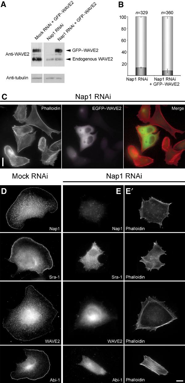
Lamellipodia formation is not restored by ectopic expression of WAVE2 in Nap1 ablated cells. Mock and Nap1 siRNA-treated B16-F1 cells with or without ectopic expression of GFP-tagged WAVE2 were analysed by Western blotting (A) or treated with AlF, fixed, stained and subjected to morphological analysis and quantification as described in Methods (B, C). Nap1 siRNA-treated cells expressing EGFP–WAVE2 were incapable of forming lamellipodia (C), although Western blotting confirmed successful expression of the GFP-tagged full-length WAVE2 protein (A). Note the significant reduction of expression of endogenous WAVE2 in Nap1 siRNA as compared to mock siRNA-treated cells (A). (B) Values are means±standard errors of means from three independent experiments. Cells were classified according to the categories (▪) with or (□) without lamellipodia. Note that ectopic expression of WAVE2 fails to increase the percentage of cells capable of lamellipodia formation (B). Measured differences between Nap1 knockdown populations with or without ectopic EGFP–WAVE expression were statistically not significant (paired t-test; P=0.11). (D–E′) Downregulation of Nap1 delocalizes Sra-1, WAVE2 and Abi-1. Mock (D) or Nap1 (E, E′) siRNA-treated B16-F1 cells were treated with AlF, fixed and stained with phalloidin (E′) or with antibodies as indicated (E). In contrast to mock-transfected controls (D), Sra-1, WAVE2 and Abi-1 were completely absent from peripheral actin structures of Nap1 siRNA-treated cells (E), which were unable to protrude lamellipodia (E′). Scale bars in (C) and (E′) equal 20 and 10 μm, respectively.