
Figure 3.
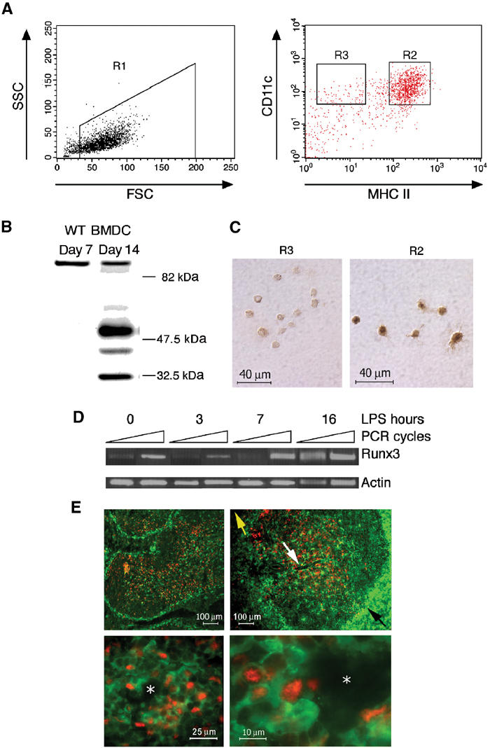
Runx3 expression is induced upon maturation of BMDC. (A) Runx3 expression in sorted WT DC. Day 11 BMDC treated with LPS (1 μg/ml), stained with anti-CD11c and anti-MHC II and analyzed by FACS. DC were gated as high forward scatter cells (R1) and sorted into CD11c+ mature, MHC II high (R2) and immature, MHC II low (R3), respectively. (B) Expression of Runx3 in spontaneously matured WT BMDC. Western blot of proteins from immature (7 days) and matured (14 days) cultured WT BMDC using anti-Runx3 Ab. Note the similar intensity of the 85 kDa nonspecific protein band in immature and mature BMDC. (C) Immunostaining of Runx3 in FACS-sorted mature BMDC. R2 or R3 cells (4 × 10³) were collected onto slides and immunostained with anti-Runx3 Ab. (D) Expression of Runx3 during BMDC maturation. Day 7 immature WT BMDC were treated with LPS (1 μg/ml), and at the indicated time points RNA was extracted and analyzed by semiquantitative RT–PCR. Equal amounts of cDNA (compared to actin) were used for PCR and the number of cycles was preoptimized. Runx3 was determined at 26 and 28 cycles (yielding a 654 bp fragment) and actin at 15 and 16 cycles (439 bp fragment) with primers indicated in Supplementary Figure S1. (E) In the spleen, Runx3 protein is detected in mature periarteriolar lymphoid sheath DC. Cryosections of spleens of CX3CR1+/GFP mice 6 h after injection of LPS (80 μg), stained with anti-GFP and anti-Runx3 Ab are shown. Upper panels are at low magnification showing the absence of Runx3 staining (red nuclear staining) in MZ immature DC (green GFP-positive cytoplasmic staining). Lower panels are at higher magnification showing Runx3 expression in mature DC located in the periarteriolar lymphoid sheaths. The yellow arrow designates the splenic red pulp, while the black designates the MZ of the white pulp. The white arrow designates the PALS surrounding the central arteriole (white asterisk).