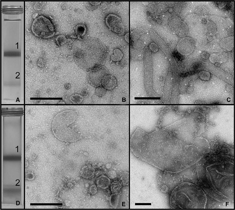
Figure 2.

Sucrose density gradient fractionation of photosynthetic membranes. (A) Result of centrifugation of cell extract from the PufX+ strain on a 15/40/50% w/w sucrose gradient. (B) Negatively stained sample from band 1 recovered from the 15/40% interface in (A). Scale bar: 0.2 μm. (C) Negatively stained sample from band 2 recovered from the 40/50% interface in (A). Scale bar: 0.5 μm. (D) Centrifugation of cell extract from the PufX− strain on a 15/40/50% w/w sucrose gradient. (E) Negatively stained sample from band 1 recovered from the 15/40% interface in (D). Scale bar: 0.2 μm. (F) Negatively stained sample from band 2 recovered from the 40/50% interface in (D). Scale bar: 0.2 μm.
