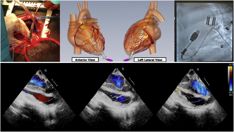
Figure 4.
Apex of left ventricle was accessed via thoracotomy. Miniaturized ventricular assist device was inserted through apex of left ventricle (top left). Graphic illustration of implanted positioning of miniaturized ventricular assist device demonstrating transapical placement (top center). Fluoroscopy shows good anatomic placement of miniaturized ventricular assist device in calf (top right). Echocardiographic color Doppler images demonstrate minimal aortic regurgitation and adequate coaptation of aortic valve with transapical outflow cannula placement (bottom).

