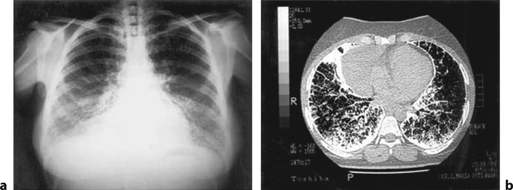
Fig. 7.
Pulmonary alveolar microlithiasis (PAM). a Chest radiograph of a patient affected by PAM, showing fine microliths with a diffuse, uniform spread obscuring the cardiac and diaphragmatic borders (sandstorm lung). b CT scan with diffuse, ground-glass opacities (stony-lung) in all pulmonary fields, and faint calcific densities, sometimes confluent at posterior and inferior subpleural regions (from fig. 1 in [54], with permission).

