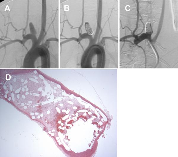
Figure 3.
A set of representative images of a single aneurysm packed with platinum coils (vitamin C pilled group). (A) Angiogram of the aneurysm cavity pre-embolization. (B) Aneurysm cavity is completely occluded immediately after coil embolization. (C) Aneurysm recanalization 12 weeks post-embolization; note that part of the aneurysm cavity has refilled with contrast, indicating recanalization. (D) Remnant neck and unorganized thrombus crossing the neck interface (H&E; magnification=12.5×).

