
Figure 2).
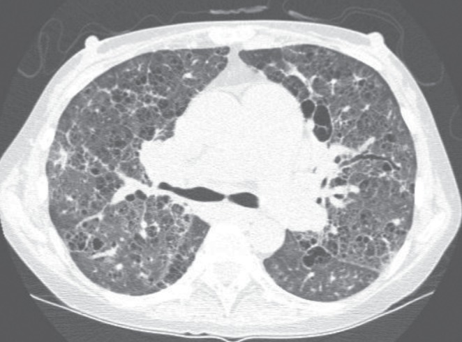
A representative section of a noncontrast computed tomography scan of the chest in patient 1. Diffuse cystic and reticular changes are apparent in addition to septal thickening throughout the pulmonary parenchyma. Bronchiectasis is also apparent