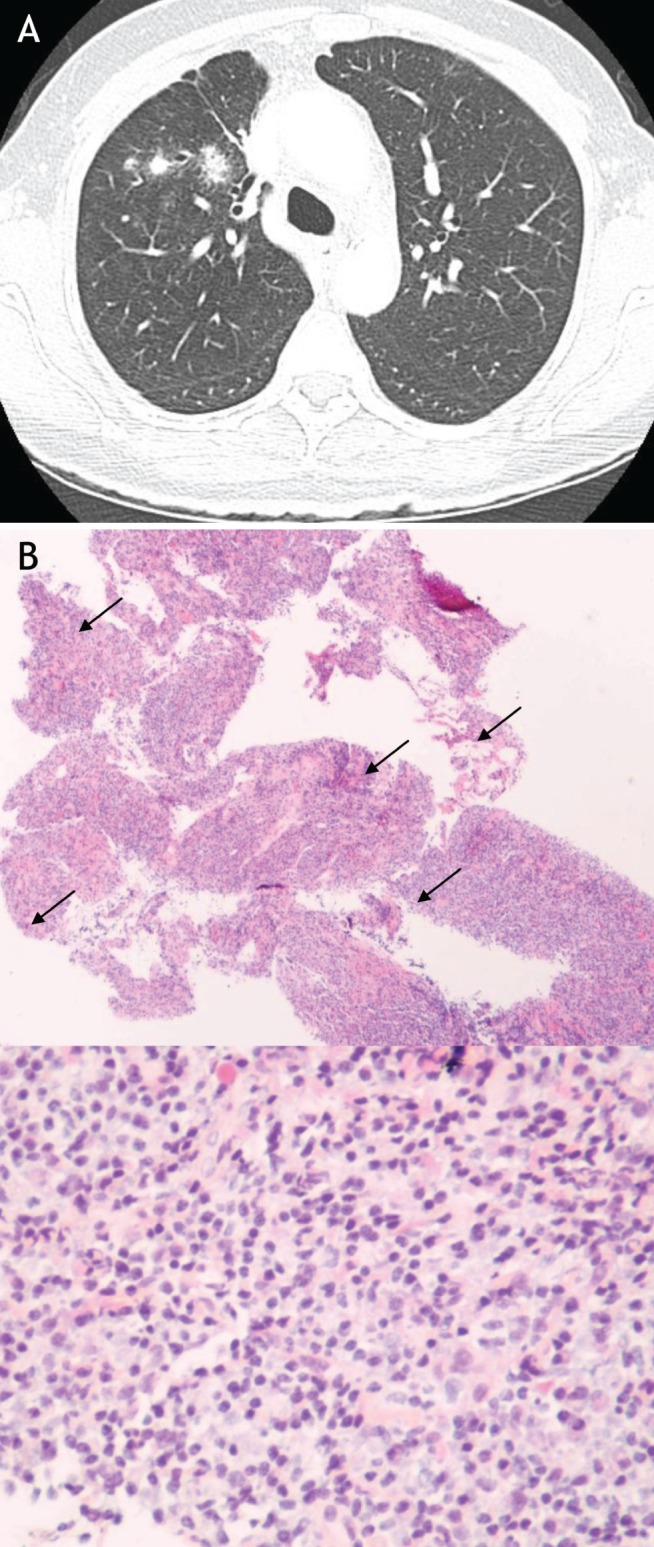
Figure 3).

A Representative section of a thoracic computed tomography scan of patient in case 2. Multiple pulmonary nodules on original computed tomography. B Histopathology of hematoxylin and eosin-stained pulmonary biopsy specimen at low- (40×) and high-power (100×) magnification. The biopsy was taken from the largest pulmonary nodule and demonstrates the characteristic fibroinflammatory infiltrate of an inflammatory pseudotumour. The high-power section illustrates the presence of numerous plasma cells (examples indicated by arrows)
