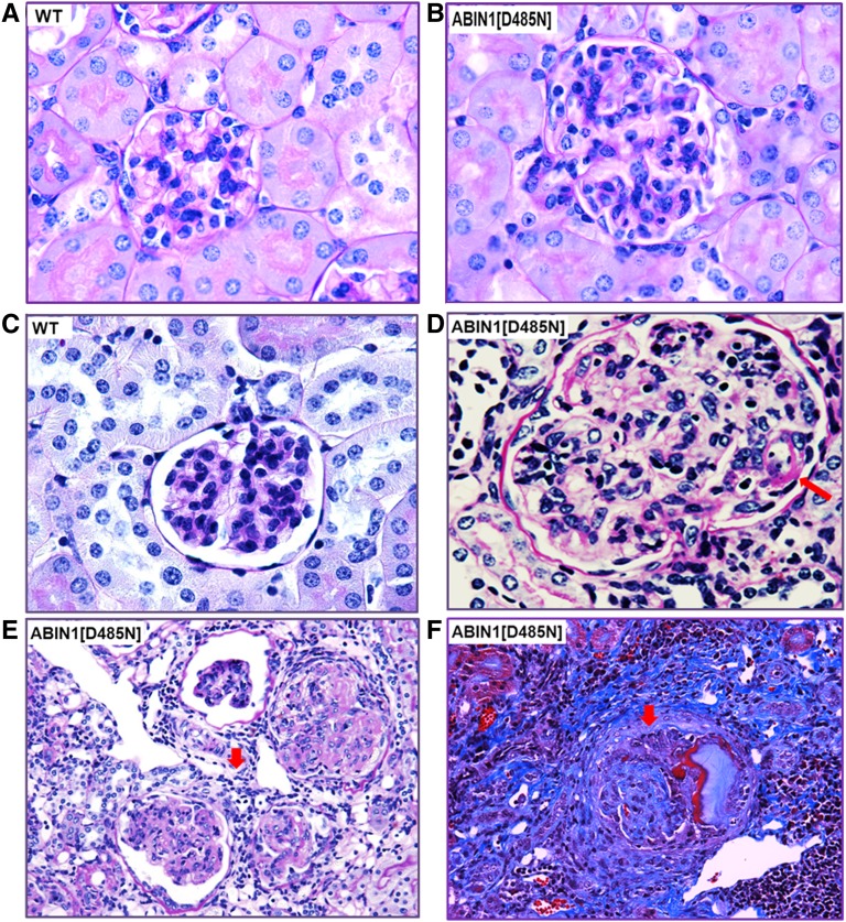
Figure 2.
ABIN1[D485N] mouse kidneys display pathologic features of proliferative immune-mediated GN. (A) 100× magnification of a periodic acid-Schiff (PAS)–stained kidney section from a 3- to 4-month-old WT mouse. (B) Comparative 100× PAS image shows that at 3–4 months, the ABIN1[D485N] mouse kidneys display mesangial hypercellularity, matrix expansion, and capillary loop thickening compared with WT mouse kidneys. (C) 100× magnification of PAS-stained kidney section from a 5- to 6-month-old WT mouse. (D) Comparative 100× PAS image shows that at 5–6 months the ABIN1[D485N] mouse kidneys display severe mesangial hypercellularity, matrix expansion, and “wire loops” (arrow) compared with WT mouse kidneys. (E) 40× magnification PAS image showing examples of glomerular injury and extensive interstitial immune cell infiltration (arrow) observed in 5- to 6-month-old ABIN1[D485N] mouse kidneys. (F) 40× magnification of Masson trichrome staining showing examples of tubulointerstitial fibrosis, glomerular fibrosis, and crescent formation (arrow) and immune cell infiltration (lower right corner) in 5- to 6-month-old ABIN1[D485N] mouse kidneys.

