
Figure 3.
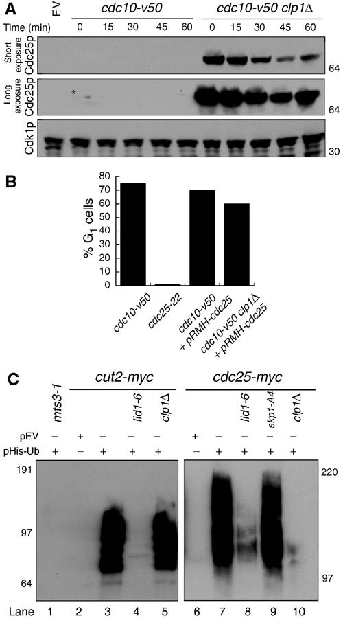
Cdc25p destabilization and ubiquitination requires Clp1p and APC/C. (A) cdc10-V50 (KGY 1744) and cdc10-V50 clp1Δ (KGY 2752) cultures expressing pREP41myc-cdc25C480S were grown in the absence of thiamine for 20 h at the permissive temperature (25°C), and then shifted to the nonpermissive temperature (36°C) for 3.5 h, at which point excess thiamine (4 μM) and cyclohexamide (100 μg/ml) were added to the cultures. Samples were taken at the indicated time points. Extracts were prepared and immunoblotted with 9E10 and anti-PSTAIR to detect Cdc25p and Cdk1p, respectively. (B) Percentage of G1 cells from (A) as determined by Sytox Green (Molecular Probes) staining and flow cytometry. (C) In vivo ubiquitination assays. mts3-1 (KGY 574) (lane 1), mts3-1 cut2-myc (KGY 1923) (lane 3), mts3-1 cut2-myc lid1-6 (KGY 1948) (lane 4), mts3-1 cut2-myc clp1Δ (KGY 878) (lane 5), mts3-1 cdc25-myc (KGY 4366) (lane 7), mts3-1 lid1-6 cdc25-myc (KGY 100) (lane 8), mts3-1 skp1-A4 cdc25-myc (KGY 102) (lane 9), and mts3-1 clp1Δ cdc25-myc (KGY 110) (lane 10) strains transformed with pREP1-His6-ubiquitin, and mts3-1 cut2-myc (KGY 1923) (lane 2) and mts3-1 cdc25-myc (KGY 4366) (lane 6) transformed with empty vector were grown at 25°C for 22 h in the absence of thiamine to induce His6-ubiquitin expression and then shifted to 36°C for an additional 4 h. Samples were collected and extracts were prepared. Ubiquitin conjugates were purified on Ni-NTA beads, separated on SDS–PAGE, and immunoblotted with 9E10 antibodies to detect Cut2p and Cdc25p.