
Figure 6.
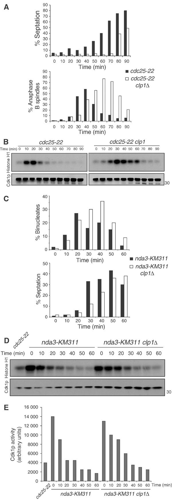
clp1Δ cells delay Cdk1p inactivation at the end of mitosis. cdc25-22 (KGY 851) and cdc25-22 clp1Δ (KGY 3380) cells were arrested in G2 by incubation at 36°C for 4 h. Cultures were then released to 25°C, and samples were taken at the indicated time points and fixed with ethanol or frozen. (A) Septation index was calculated using a light microscope (top panel). Ethanol-fixed samples were subjected to indirect immunofluorescence with anti-TAT1 antibodies (bottom panel). The presence of elongated spindles and separated DNA masses was assessed in at least 200 cells. (B) Cell pellets were lysed under native conditions and immunoprecipitated for Cdk1p with 4711 antibody. Samples were processed for histone H1 kinase activity and immunoblotted for Cdk1p levels with anti-PSTAIR. (C) nda3-KM311 (KGY 3612) and nda3-KM311 clp1Δ (KGY 3783) were arrested in prometaphase by incubation at 18°C for 6.5 h. Cells were released at 32°C, and ethanol-fixed samples and cell pellets were collected at the indicated time points. Septation index was determined as in (A) (C, lower panel) and fixed cells were stained with DAPI to visualize nuclei. The presence of separated DNA masses and no septum was assessed in at least 200 cells (C, top panel). (D) Cell pellets were analyzed as in (B) and Cdk1p activity was quantified (E).