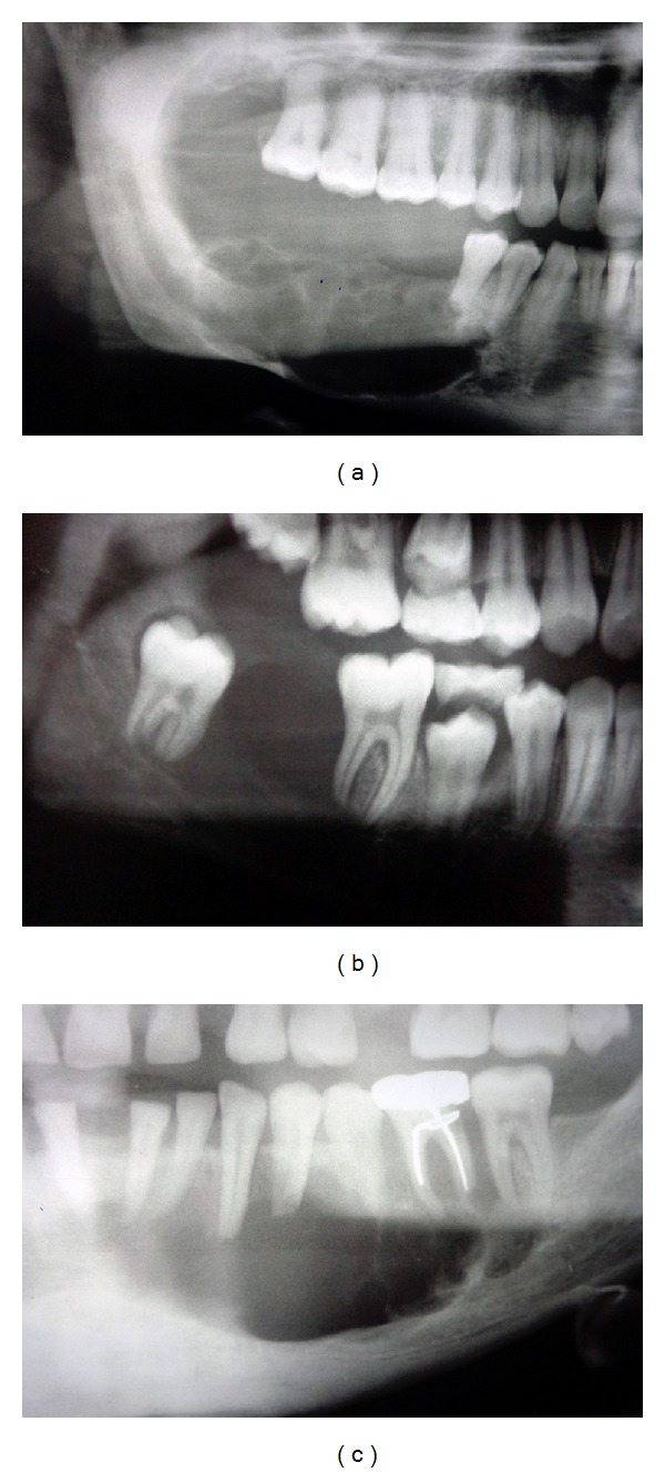
Figure 1.

Radiographic presentations of unicystic ameloblastoma: (a) multilocular radiolucency with scalloped border, cortical thinning, perforation, and resorption; (b) well-defined unilocular radiolucency; (c) multilocular radiolucency with root resorption.
