
Figure 6.
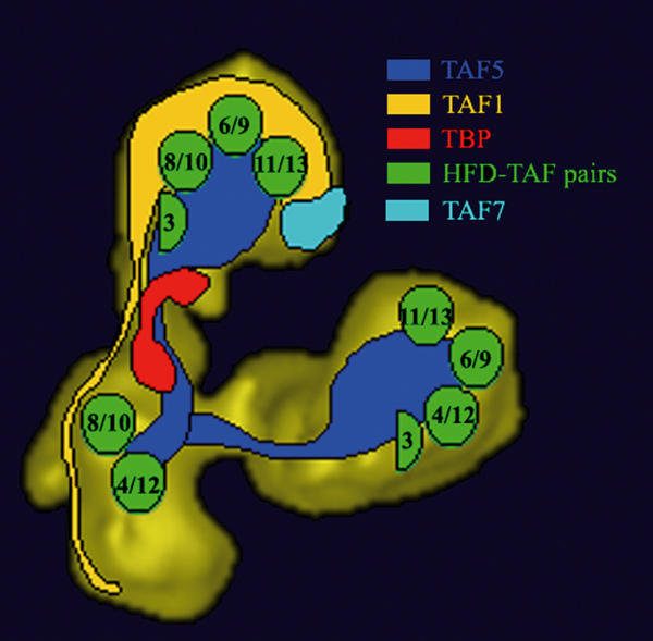
Summary of the immunolabelling experiments. Proposed location of TAFs within the 3-D model of yTFIID. The location of histone fold-containing TAF pairs is schematically represented by green circles as determined previously (Leurent et al, 2002). The extended TAF1 and TAF5 subunits are schematically represented to highlight their presence in two lobes, Note that the size and shape of the coloured areas does not directly correspond to polypeptide mass.