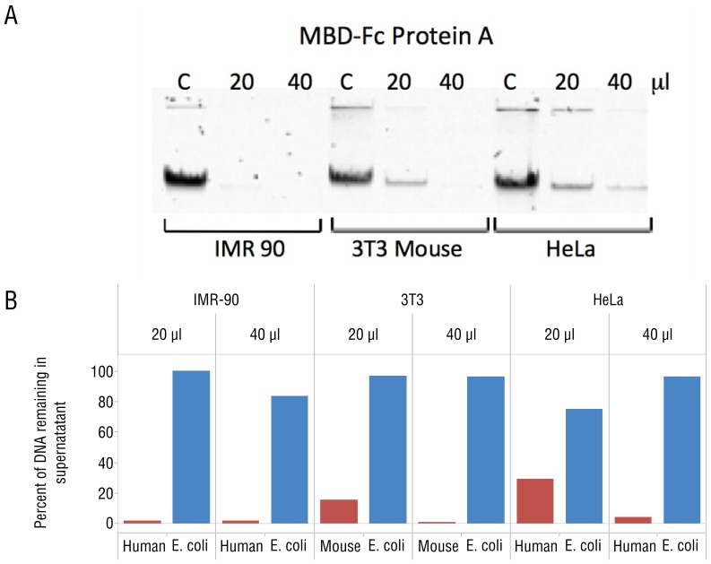
Figure 2. MBD-Fc fusions bind mammalian DNA.
(A) Gel image demonstrating depletions of mammalian DNA by MBD-Fc binding. A total amount of 250 ng input DNA was incubated with increasing amounts of MBD-Fc beads as indicated above the gel. “C” corresponds to a control reaction with no MBD-Fc beads in the incubation mixture. The unbound, supernatant fraction of each mixture was resolved and is shown on the gel image. (B) Separation and quantitation of bound (mammalian) and unbound (E. coli) DNA in a mixture containing DNA from both sources. Gel densitometry results are shown with increasing bead quantity in the depletion experiment. The percentage of E. coli DNA in the supernatant was calculated by 3H scintillation counting and comparing with the input counts per minute.

