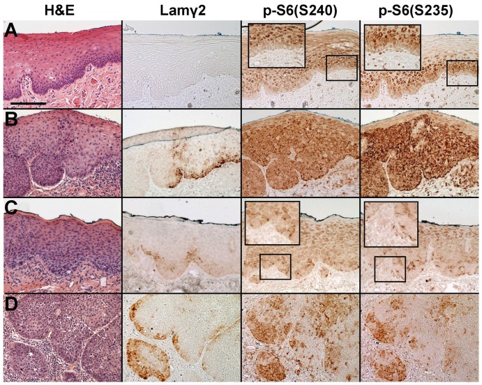
Figure 2. Coincidence of p-S6(S235) and p-S6(S240) detectable in human oral dysplastic lesions and SCCs in vivo.
Sections of normal (A) and dysplastic (B-C) epithelium, and invasive SCC (D), stained with H&E and immunostained for Lamγ2, p-S6(S240) and p-S6(S235). Scale bar: 200 µm. Enlarged insets of some regions are shown for easier viewing of p-S6 staining patterns. Panel A shows a region of Case 2, panel B of case 8, panel C of case 7, and panel D of case 4 as described in Table 1. Note that normal epithelium did not express Lamγ2 and neither S6 phosphorylation event was detectable in the basal cells. Cells in dysplasias and SCCs always showed coincidence of the two S6 phosphorylation events. Dysplasias varied with respect to frequency and intensity of Lamγ2 expression and S6 phosphorylation, with Lamγ2 cells representing a subset of p-S6 positive basal cells and invasive SCCs contained many Lamγ2 and basal layer p-S6 positive regions.

