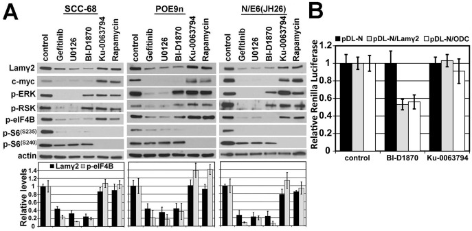
Figure 3. MAPK/RSK-dependent, mTOR/S6K1-independent activation of eIF4B and Lamγ2 mRNA translation.
A) Western blot analysis of confluent cultures of SCC-68, the premalignant oral keratinocyte line POE9n, and normal primary keratinocyte strain N engineered to express the JH26 mutant of HPV16 E6 (N/E6(JH26). Cultures were treated for 24 hr with the indicated kinase inhibitors and then analyzed for levels of Lamγ2 and MYC protein and for the phosphorylated, activated forms of signaling proteins and translation factors. The Lamγ2 band shown is the 155 kD intracellular form and not the 105 kD form that predominates after secretion and proteolytic processing. The bar graphs below show densitometric analysis of Lamγ2 and p-EIF4B levels in each drug treatment condition relative to untreated control cultures of each line, as described in panel B. B) SCC-13 cells transfected with the reporter constructs pDL-N, pDL-N/(Lamγ2 5′-UTR), and pDL-N/(ODC 5-′UTR) with or without the RSK inhibitor BI-D1870 or the mTORC1/2 inhibitor Ku-0063794 and analyzed for Renilla and Firefly luciferase activity. Reduction caused by BI-D1780 in Lamγ2 5′UTR- and ODC 5′UTR-dependent expression had P values for significance of 0.0043 and 0.01, respectively.

