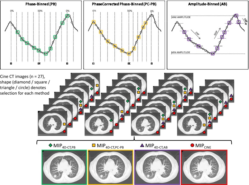
Figure 1.
MIP images generated from one of the three 4D-CT sorting processes (PB, PC-PB, and AB) or from the use of all 27 cine CT images. Top: Respiratory signal at a single bed position on a representative patient case showing that the same input will lead to the selection of a different distribution of ten images for each different sorting method (bottom). Each of the three image sets is generated from the selected images.

