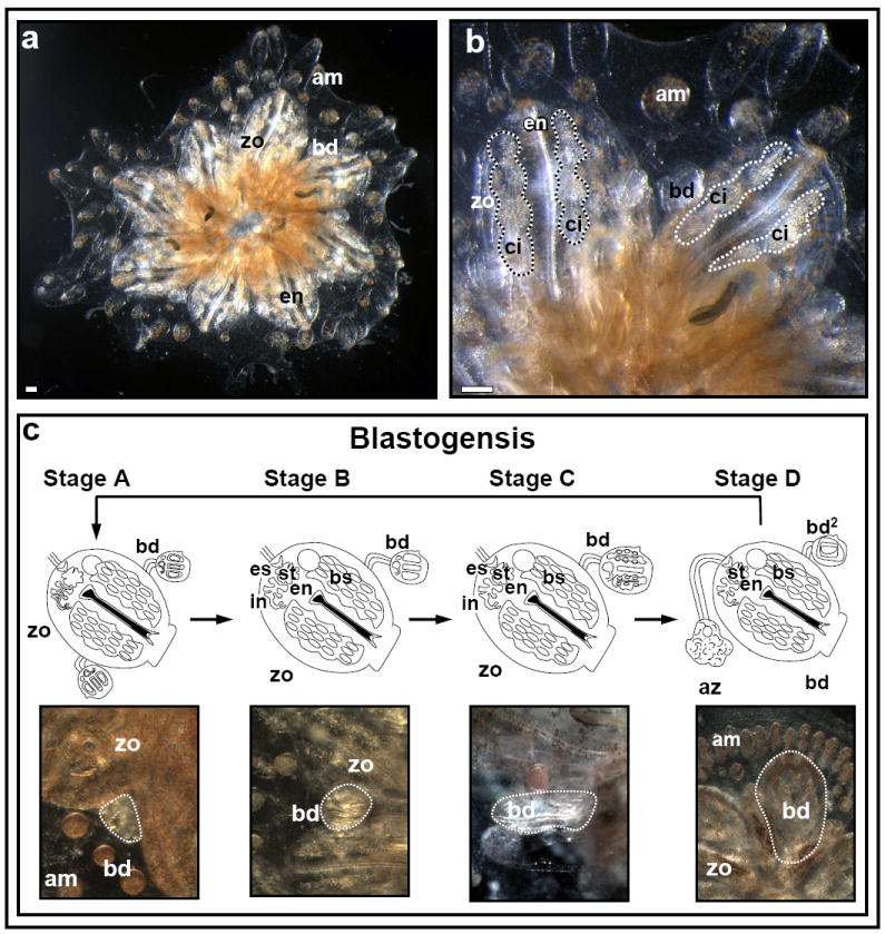
Figure 1. The anatomy and developmental cycle (blastogenesis) of Botryllus schlosseri.

(A). A Botryllus schlosseri colony composed of a system of eight zooids (zo) and developing buds (bd), fringed at the periphery by blood-vessel termini called ampullae (am). (B) High power magnification of a Botryllus schlosseri colony. Each adult zooid houses a feeding branchial grove, termed endostyle (en), along with several lateral blood cell aggregates (termed ‘cell-islands’; ci) within CI niches. (C) Colonies of botryllid ascidians grow through weekly cycles of development termed blastogenesis. Each blastogenic cycle is composed of four major stages (A-D), during which, new buds emerge from the body wall of each parental zooid. Each blastogenic cycle ends in a massive apoptotic and phagocytic event of all parental zooids concurrent with fast development of primary buds to the adult zooid stage (stage D, also called ‘takeover’). Scale bars=100μm.
