
Figure 3. Putative stem cell migrations to developing CIs and the germ-line.
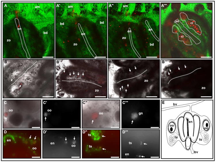
Fate of labeled EN cells: (A-A’”, the endostyle is outlined by a dotted line). Representative Vybrant DiD labeling (665nm emission spectra) of 20-60 EN cells (red) of a colony at blastogenic stage B (A). Labeled cells proliferate within the inter-zooid sinuses and migrate to developing buds during blastogenic stage B (A’) and throughout stage C (A”). Mature primary bud replacing the old generation zooid on blastogenic stage D; labeled cells are detected within the new generation of zooids sinuses and in CIs lateral to the endostyle (A’”, red; CIs: dotted circle). Fate of labeled CI cells (B-B’”, white arrows). Representative label of 10-50 cells from the anterior CI, within a colony at blastogenic stage B (B, white arrows). During blastogenic stages B,C, labeled cells appear within posterior CIs and on both lateral sides within the same zooid (B’, B”; white arrows; two representative zooids 3 days following CI labeling). Following ‘take-over’ labeled cells reappear in newly developed CIs (B’”, white arrows; 6 days following CI labeling). Labeled oocytes (C-C’”, D, D’, white arrows) and testis (D”, D’”, white arrows) within the gonads of a subsequent generation, 10 days following initial label. Dotted line in B-B’” outlines the endostyle groove. (E) Schematic illustration of the zooid anatomy as seen in cross sections in C and D. am, ampullae; bd, primary bud; bv, blood vessel; ci, cell island; en, endostyle; oo, oocyte; ts, testis; zo, zooid. A-C’” in vivo imaging; D-D’” frozen sections. Longer exposure times were used to detect and image the descendent of the EN / CI Vybrant DiD labeled cells A’-A’” and B’- C’” compare to A and B. Scale bars=100μm. See also Figure S2.