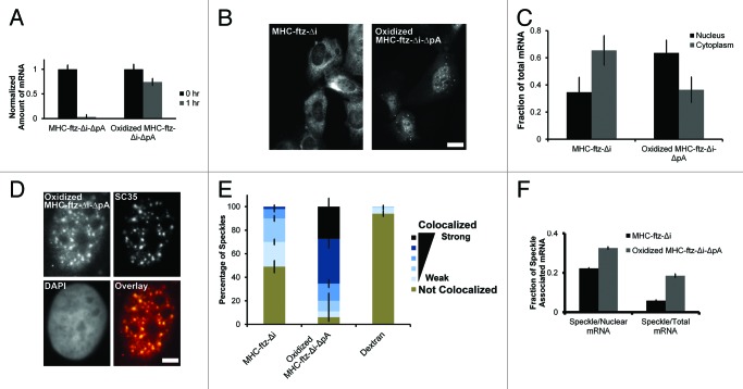
Figure 7.MHC-ftz-Δi mRNA lacking a poly(A)-tail accumulates in nuclear speckles and is poorly exported. (A) In vitro transcribed MHC-ftz-Δi mRNA lacking a poly(A)-tail (ΔpA) was either oxidized with periodate or left untreated. These mRNAs were microinjected into the nuclei of human U2OS cells, which were then immediately fixed (“0 hr”) or first incubated for 1 h at 37°C then fixed. Cells were probed for ftz and imaged. The total mRNA fluorescence was quantified for each nuclear-injected cell. Each bar represents the average and standard error of 12‒30 cells, all values being normalized to the average fluorescent intensity at 0 h. (B) Nuclei of cells were microinjected with either in vitro transcribed and polyadenylated MHC-ftz-Δi mRNA or with periodate oxidized MHC-ftz-Δi-ΔpA. After allowing mRNA export to proceed for 1 h, cells were fixed, stained using FISH, and imaged. Scale bar = 20 µm. (C) The fraction of mRNA in the cytoplasm and nucleus was quantified. Each bar represents the average and standard error of three independent experiments, each consisting of 12‒30 cells. (D) An example of a nucleus microinjected with in vitro transcribed and oxidized MHC-ftz-Δi-ΔpA mRNA and then incubated for 1 h before fixed, probed for ftz mRNA and immunostained for the nuclear speckle marker SC35. An overlay of the mRNA (red) and SC35 (green) is shown in the bottom right panel. Scale bar = 5 µm. (E) The percentage of nuclear speckles that demonstrate different levels of colocalization with MHC-ftz-Δi, periodate oxidized MHC-ftz-Δi-ΔpA and dextran 1 h after mRNA microinjection. Data was analyzed and plotted as described in Figure 1D. (F) The percentage of total cellular and nuclear MHC-ftz-Δi and periodate oxidized MHC-ftz-Δi-ΔpA mRNA that is present in nuclear speckles as described in Figure 1H.

An official website of the United States government
Here's how you know
Official websites use .gov
A
.gov website belongs to an official
government organization in the United States.
Secure .gov websites use HTTPS
A lock (
) or https:// means you've safely
connected to the .gov website. Share sensitive
information only on official, secure websites.
