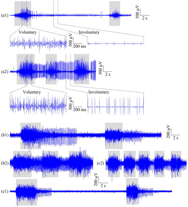
Figure 3.
Illustration of representative signal segments of a single surface EMG channel recorded from (a) Subject 3, (b) Subject 6, and (c) Subject 8, within (1) the slow session and (2) the fast session, respectively, when the subject was performing cylindrical grip. The gray rectangles under every signal segment mark voluntary muscle contractions. Each of the top two signal segments, (a1) and (a2), is also shown with an overview (top) and two expanded views (bottom) of voluntary and involuntary EMG activity, respectively.

