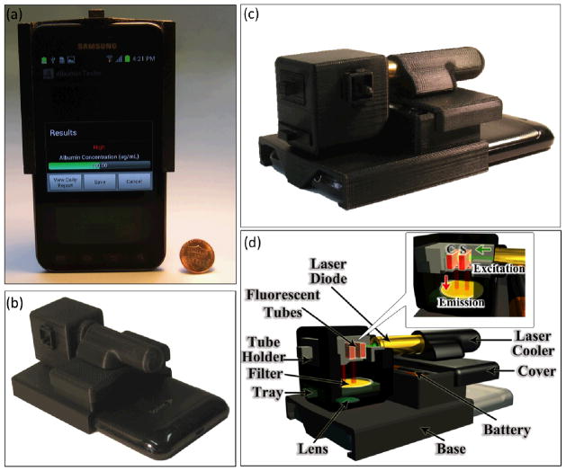
Fig. 1.
(a–c) Photographs of the Albumin Tester installed on a smart-phone, weighing 148 grams, are shown from different views. (d) Schematic diagram of the lightweight smart-phone attachment of the Albumin Tester is illustrated, where the test and control tubes are inserted from the side and are pumped by a battery powered laser diode. After probing the sample of interest located within the test tube, this excitation beam interacts with the control tube. The fluorescent emission is then collected perpendicular to the direction of excitation, where the cellphone camera captures the images of the fluorescent tubes through the use of a simple plastic lens that is inserted between the tubes and the cellphone camera lens. These acquired raw fluorescent images of the sample and control tubes are digitally processed within 1 s using a custom-developed Android application running on the same smart-phone for detection and quantification of albumin concentration in the urine specimen.

