
Figure 1.
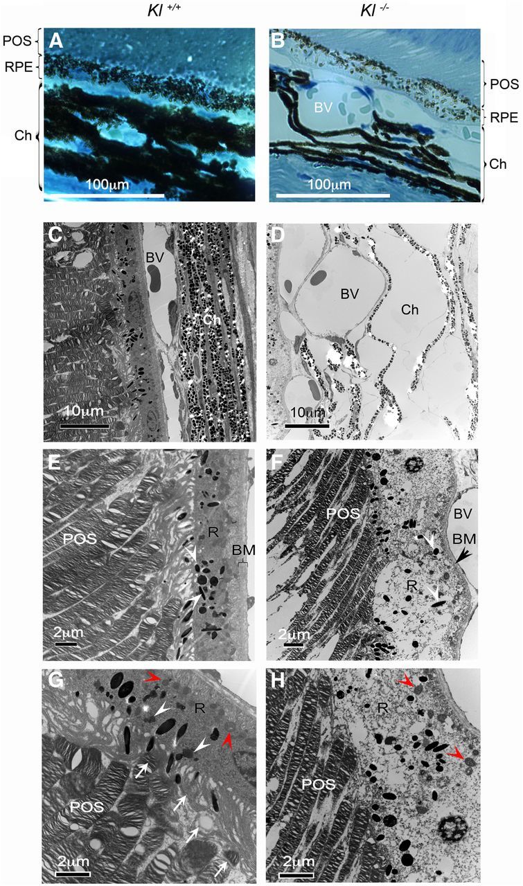
Klotho knock-out mice exhibit degenerative phenotypes in the retina. A, B, Light microscopy images of the retina of adult Kl+/+ and Kl−/− mice. The number of pigmentation granules in RPE cells is reduced by 30%. Eyes from four 6-week-old Kl−/− (B) and four age-matched Kl+/+ (A) mice were dissected for histological analysis. The number of melanin granules was counted in 10 RPE cells from each mouse; the statistical analysis of the results is presented in Table 1. The choroid (Ch) of the Kl−/− mice showed deformed layers separated by dilated blood vessels (BV; B). POS, Photoreceptor outer segments. C–H, Electron microscopy images of the retinal region of adult Kl+/+ and Kl−/− mice: choroid, RPE, and outer segments of the photoreceptors of wild-type mice (C); choroid and RPE of Kl−/− mice (D; the choroidal region appears grossly deformed with tissue layers that have been separated by severely dilated BVs; RPE (R) of Kl+/+ mice, showing normal cytoplasmic content, normal mitochondria, and Bruch's membrane (E); the RPE (R) of Kl−/− mice are degenerated with light cytoplasmic content, damaged mitochondria (H, red arrowheads), and melanosomes with disorganized distribution (F, white arrowheads). BM is thinner, deformed by dilated blood vessels, showing significant degeneration; F, black arrowhead). The POSs are thinner, showing signs of degeneration (F, H) compared with the Kl+/+ photoreceptors (E, G); phagocytosis of the POSs is observed in the RPE cells of Kl+/+ mice (G); arrows indicate presumptive phagocytotic vesicles at different stages of the phagocytosis process. White arrowheads show the mature lysosomes. Red arrowheads show normal mitochondria. H, No signs of phagocytosis of the outer segments of photoreceptors were observed in the RPE cells of Kl−/− mice. Red arrowheads show damaged mitochondria.