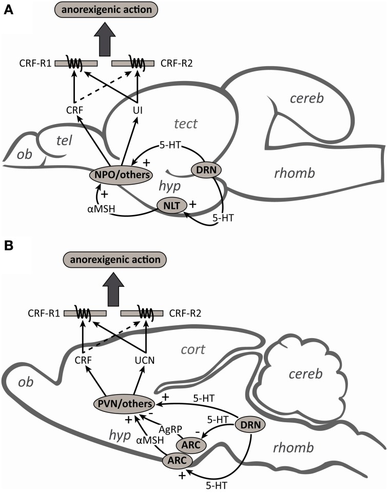
Figure 6.
Schematic diagrams of midsagittal sections through (A) the rainbow trout and (B) rat brains to summarize the contributions and interactions of the serotonergic and corticotropin-releasing factor (CRF) systems in the regulation of food intake. Serotonergic (5-HT) neurons from the dorsal raphe nucleus (DRN) can directly stimulate the release of CRF from the nucleus preoptic (NPO) and paraventricular nucleus (PVN) in fish and mammals, respectively. In mammals, 5-HT neurons also activate arcuate nucleus (ARC) neurons to facilitate the release of the melanocortin 4 receptor (MC4R) agonist α-melanocyte-stimulating hormone (αMSH) and inhibit the release of the MC4R antagonist agouti-related peptide (AgRP). CRF neurons in the PVN act as a downstream mediator of MC4R signaling and contribute to the regulation of food intake. In fish, while the anorexigenic actions of αMSH also appear to be mediated by the CRF-signaling pathway, the interactions between 5-HT and the melanocortin system have yet to be investigated. Also unknown are the roles of urotensin I (UI) and urocortin (UCN) secreting neurons as mediators of either the direct or indirect anorexigenic actions of 5-HT. Although CRF and UI/UCN have anorexigenic actions and both the CRF type 1 (CRF-R1) and type 2 (CRF-R2) receptors have been implicated in the regulation of food intake, the higher affinity of UI/UCN than CRF for CRF-R2 likely explains why UI/UCN more potently inhibits food intake than CRF (see text for further details). Abbreviations: cereb, cerebellum; cort, cerebral cortex; hyp, hypothalamus; NLT, nucleus lateralis tuberis; ob, olfactory bulb; rhomb, rhombencephalon; tect, optic tectum; tel, telencephalon.

