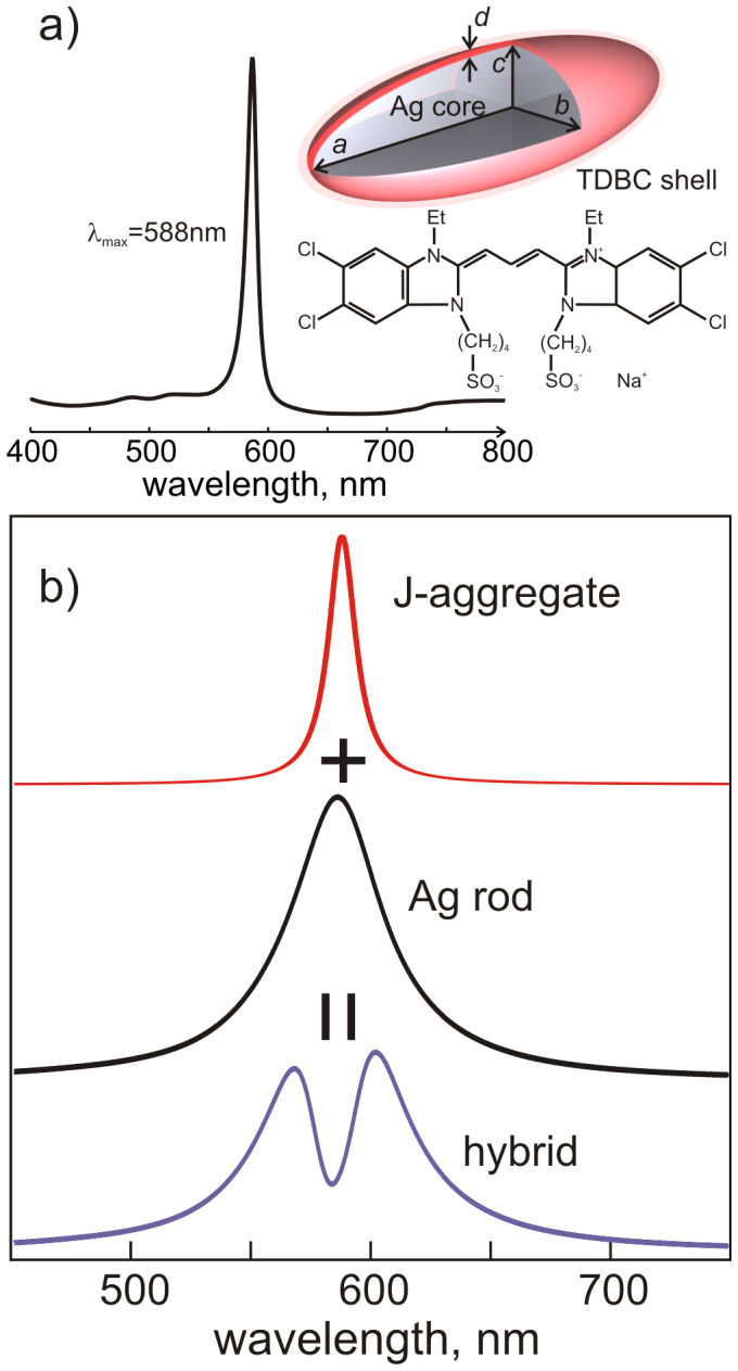
Figure 1.

(a) A sketch showing the Ag core – molecular shell spheroid with semiaxes a, b, c, uniform shell thickness – d and the chemical structure of TDBC. Inset: the absorption spectrum of 10−5 M TDBC J-aggregate in aqueous solution containing 5 mM NaOH. (b) Absorption spectrum of free J-aggregate (red) and scattering spectrum of a bare Ag nanorod (black), which upon interaction, give rise to a hybrid plasmon-molecule spectrum (purple).
