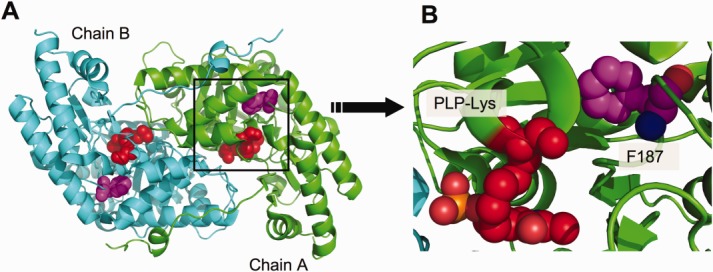
Figure 1.

(a) Cartoon representation of the S187F variant dimer. The mutated amino acid (magenta) and the PLP-binding lysine (red) is represented by spheres. (b) Magnification of the area around the PLP-binding lysine. Because of the position of the phenylalanine mutation relative to the co-factor binding site, a conformational change is induced in the close proximity of the PLP-binding lysine. [Color figure can be viewed in the online issue, which is available at wileyonlinelibrary.com.]
