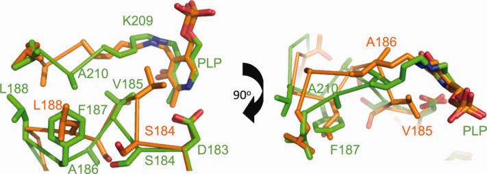
Figure 2.

Superposition of the structure of the S187F variant (green) with that of normal AGT (orange) in the area of the cofactor binding site. Residues are labeled with the respective colors. The hydrophobic cleft that is formed by residues 185-188 triggers a conformational change in the loop where the PLP-binding lysine is located. [Color figure can be viewed in the online issue, which is available at wileyonlinelibrary.com.]
