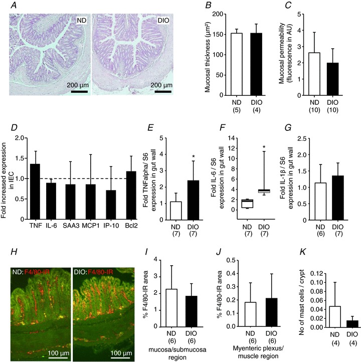
Figure 2. Normal appearance, integrity and permeability of distal colon in DIO mice (12 weeks of feeding).

Haematoxylin and eosin staining revealed normal histology of the distal colon in DIO mice (A) and no increased mucosal thickness (B). Paracellular permeability was comparable between ND and DIO mice as mucosal to serosal translocation of fluorescein sulfonic acid was unchanged (C). The expression profile of markers for inflammation [tumour necrosis factor (TNF), interleukin-6 (IL-6), serum amyloid A 3 (SAA3)], chemotaxis [monocyte chemoattractant protein 1 (MCP1), interferon-inducible protein-10 (IP-10)] or apoptosis (B-cell lymphoma 2, Bcl2) was similar in ND (six animals) and DIO (four animals) mice (D). However, in extracts of the entire colonic wall (including fat tissue), the expression of TNF-α (E) and IL-6 (F), but not interleukin-1β (IL-1β) (G), was increased in DIO mice. Images in (H) are representative examples of F4/80 immunoreactive macrophages in cross-sections of the distal colon in ND and DIO mice. Analysis of macrophage densities in the mucosa/submucosa region (I) and myenteric plexus/muscle layer region (J) revealed no increased macrophage density in either region in DIO mice. Likewise, the number of CD117-positive mast cells was not altered significantly (K). Numbers in parentheses indicate number of animals. Asterisks mark significant changes, see text for P values. DIO, diet-induced obese mice; IEC, isolated epithelial cell; ND, mice receiving normal diet.
